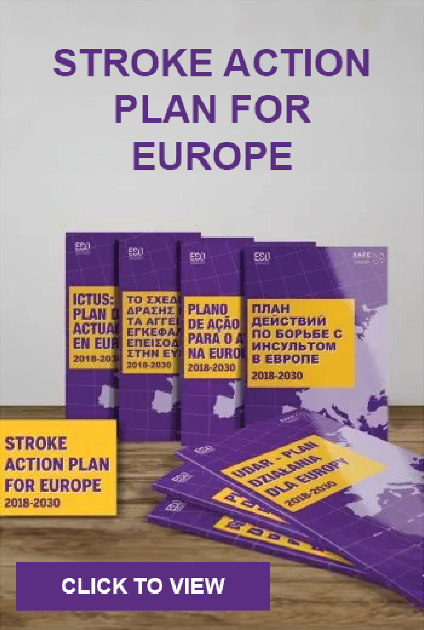
Feb 27, 2020
Slovak weekly magazine in cooperation with RTVS -Slovak national TV and Radio and with the Slovak National Theater presented the nominees of the twelfth year of the SLOVENKA OF THE YEAR.
JUDr. Alžbeta Husarovič, MHA, MPH is one of this year’s laureates for the Slovakian Women of the Year. She is the President of the civic association Porážka.sk, which is dedicated to raising stroke awareness and helping patients and their families with information about physiotherapy, speech therapy, psychology, social assistance. The aim of this organisation is to improve patients’ quality of life and lead them to self-sufficiency.
“I was very pleased and amazed when I became the Nominee for the Slovak Woman of the year 2020. I didn’t think that somebody is even noticing me for what I am doing. And I am doing this activity with love” said Alžbeta, adding: “This event Slovak Woman is a competition but I don’t consider it so. For me, it is a opportunity for woman to show others what the can do!”
The Official Press Release “Slovakia 2020”
We have no idea yet of which successful Slovak woman will be awarded in the following categories: Business and Management, Arts and Culture, Media and Communication, Science and Research, Education, Support for Young Talents, Healthcare, Sport and Charity, since all laureates are inspirational ladies, who will be introduced to you over the next few weeks.
In addition, this year the nomination committee will award the title of Absolute Slovak Woman of the Year and the Special Lifetime Achievement Award for Slovakia. We will come to learn the winning names during the gala evening in the historical building of the Slovak National Theater and live broadcast of RTVS – Slovak National TV and Radio on May 31, 2020.
Throwback
Historically, the first Slovak Woman of the year in 2009 became the head physician of the 2nd Pediatric Clinic of the Faculty of Medicine and the Pediatric University Hospital with the Policlinic in Bratislava MUDr. Anna Hlavatá, and so far the last – Slovak of the year 2019 – is Andrea Gontkovičová.
Voting from today at www.slovenkaroka.sk
Starting today, it is possible to vote for individual nominees at www.slovenkaroka.sk and from next week also in the weekly Magazine Slovenka. Vote for your favorite and win an exclusive 8-day vacation stay for 2 people at Labranda Coral Beach 4 * with all inclusive in attractive Gambia from CK Hydrotour.
<End>
Image credits: Porazka.sk
Feb 25, 2020
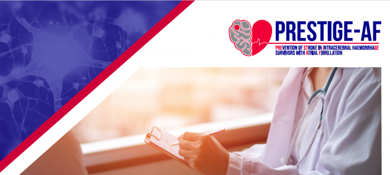
Stroke is one of the largest public health challenges around the world, and occur when the blood supply to the brain is interrupted, such as through a clot or a brain bleed. It is the most common cause of adult-acquired disability, the second leading cause of death globally and the second most frequent cause of dementia. In addition, its impact is expected to further increase in the coming decades due to the ageing population.

Dr Eleni Korompoki, MD, PhD, FESO, PRESTIGE AF researcher
We spoke with Dr Eleni Korompoki, MD, PhD, FESO, about a €6.9m worth EU funded research project, aimed at patients with atrial fibrillation (AF), a common heart condition which causes irregular and abnormally fast heartbeat, who have previously had a stroke caused by bleeding in the brain (termed intracerebral hemorrhage or ICH). The Prevention of Stroke in intracerebral hemorrhage survivor with Atrial Fibrillation (PRESTIGE-AF) brings together scientists and clinicians across Europe with the goal of reducing the risk of further stroke in this group of patients. Dr Korompoki is a Clinical Research Fellow in Stroke Medicine at Division of Brain Sciences, Imperial College London.
SAFE: If you were to explain the project’s aim to a person without any medical background, what would you say?
EK: We are conducting a research study to show whether or not patients who have had a brain hemorrhage should be given anticoagulant medication to prevent blood clots that can be caused by a heart arrhythmia called atrial fibrillation. Currently we do not know the best way to prevent strokes in these patients.
SAFE: What types of partner do you need to carry out a project like this?
EK: This project is covering many specialties, so we need to have partners with experts in stroke, cardiology, genetics, biomarkers, neuroimaging and predictive modelling to name just a few!
SAFE: Can you briefly describe your role in the project?
EK: I am part of the central clinical trial team who assist all of the (70!) hospitals who recruit to our clinical trial. I am available to provide medical support, answer questions about patient eligibility and review the safety reports (called adverse events) as we closely monitor all health complaints that patients have whilst they are in the study. Towards the end of the project I will be involved with data analysis and writing up the results of the trial.
SAFE: What (if any) are the difficulties with carrying out the work?
EK: Stroke is the leading cause of disability in Europe and strokes caused by bleeding tend to be more disabling. Therefore it can be a challenge to identify patients who are willing and able to take part in a study like this.
SAFE: What personally attracted you to be in this project?
EK: I have been involved in stroke research for more than 10 years, focusing on stroke prevention and heart-brain connection. As a physician and researcher, I strongly believe that prevention is the key element for well- being. PRESTIGE-AF is a prevention trial targeting an individualised approach and better quality of life after stroke. The trial will also address important aspects such as gender differences, and patients’ attitudes and preferences.
As part of PRESTIGE-AF consortium I have plenty opportunity to interact with internationally recognised experts of ten leading European academic institutions, to work together with a multidisciplinary team from different European countries gaining a lot of experience and improving my scientific skills.
SAFE: When this project ends, what do you expect to change, i.e. how it will reflect on stroke treatment?
EK: We expect to be able to provide evidence based treatment to patients who have had a brain hemorrhage and have atrial fibrillation, the most common cardiac arrhythmia. In addition we hope that we will be able to start using a more person centered approach with these patients using information learnt from looking at brain scans, blood tests, gender differences and psychological aspects.
PRESTIGE-AF has received funding from the European Union’s Horizon 2020 research and innovation programme under grant agreement No. 754517.
Feb 20, 2020
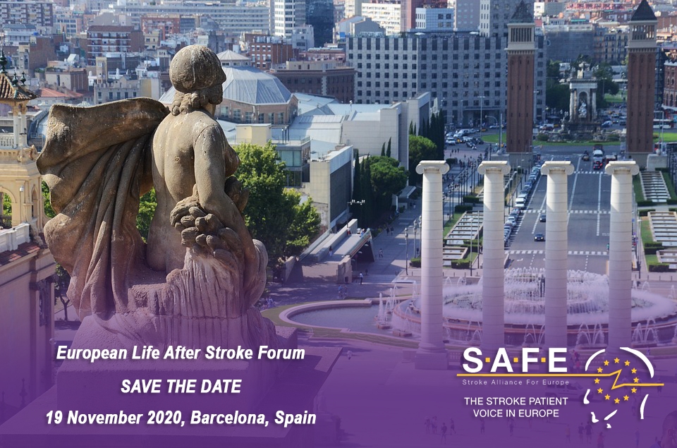
Life after stroke is a key priority within SAFE’s strategy. It is our pleasure to share with you the date of the first European Life After Stroke Forum – the 19 November 2020 in Barcelona, Spain.
This first European Life After Stroke Forum is driven by the need to implement the Stroke Action Plan for Europe and to create a network of stakeholders involved in professionally researching, advocating and providing evidence for improved life after stroke care.
SAVE THE DATE – EUROPEAN LIFE AFTER STROKE FORUM, 19 NOVEMBER 2020, BARCELONA, SPAIN
We hope we could get you to support the European Life After Stroke Forum by sharing this information with people you know.
When? 19 November 2020
Where? Hotel Catalonia Barcelona Plaza, Barcelona, Spain
Who can attend? Organisations and individuals who operate in the life after stroke area and are research, policy, advocacy or support oriented.
How to register? The registration link will be available soon. Stay tuned!
Please put this date in your calendar and stay tuned for more information that will follow.
We hope to welcome you to Barcelona,
SAFE team
Feb 20, 2020
Turning the Tide is a series of 26 mini-films from 24 countries, presenting the bold actions being carried out by communities and organisations to take on the world’s biggest killers – non-communicable diseases, like cancer, diabetes and lung disease – and bring better health for all.
Time is everything when dealing with stroke patients, but many hospitals are not “stroke ready”. Every 30 minutes a stroke patient who could have been saved, dies or is permanently disabled, because they were treated in the wrong hospital.
The Angels Initiative is building a global community of stroke centres and stroke-ready hospitals, working every day to improve the quality of treatment for every stroke patient. The story in this video would have been very different if it wasn’t for a joint effort of the Angels Community.
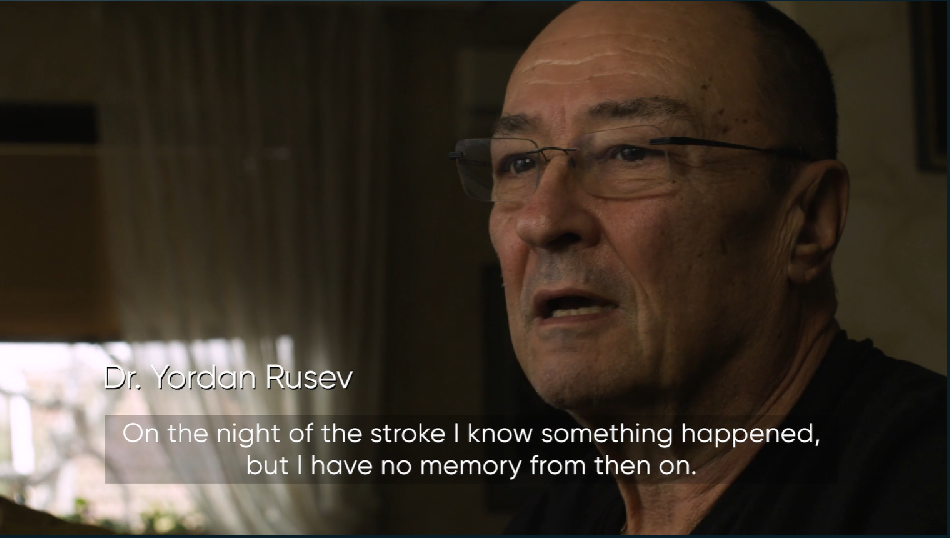
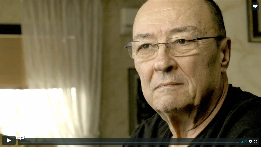
The man who can be seen in the video is one of the lucky ones. Both he and his wife are medics, so, when he collapsed from a stroke in his home in Sofia, Bulgaria, his wife instantly recognised what happened and knew that she had to act quickly. He was swiftly taken to the nearest stroke-ready hospital and was treated immediately by the responding doctors. Thanks to a swift reaction from their colleagues, medics who work in the stroke-ready hospital and to the timely received treatment, the man was able to recover. “I started to go shopping. I started driving. I started to feel like a normal human being again. These days I realise how blessed I was to have fully recovered to be with my family, my children and my grandchildren” he said.
Please watch the video and see how having stroke-ready hospitals helps #TurningTheTide.
The video was produced by the NCD Alliance and BBC StoryWorks
The Angels Initiative, launched by Boehringer Ingelheim in partnership with the European Stroke Organisation (ESO), and the World Stroke Organisation (WSO), the Stroke Alliance for Europe (SAFE) and others is set to improve acute stroke care, by increasing the number of patients treated in stroke ready hospitals and optimising the quality of treatment in all existing stroke centres. For more information about Angels Initiative, please visit www.angels-initiative.com
Image source: The video print screen
Feb 19, 2020
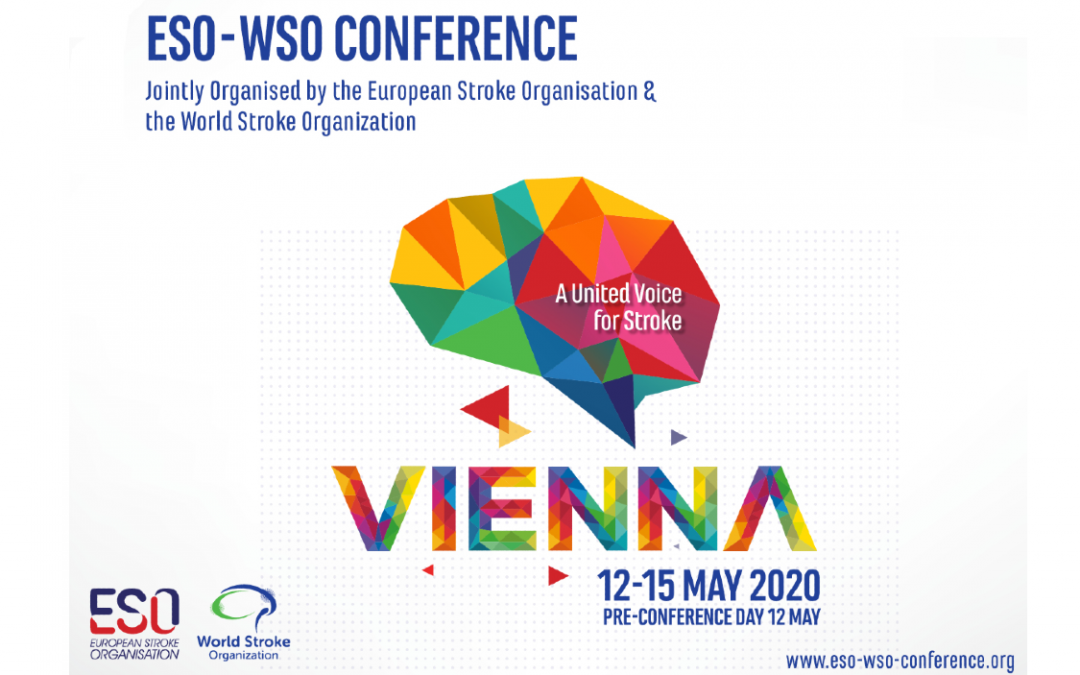
ESO-WSO 2020 congress is fast approaching. For the first time, two large congresses, the World Stroke Congress and the European Stroke Congress will merge into one, bringing together stroke researchers, medical experts, patient organisations, pharma industry, medical devices manufacturers and many others to Vienna, Austria between 12 – 15 May 2020.
The Stroke Alliance for Europe will have a stand at this big event, seizing opportunity for a direct communication with the congress delegates and promoting our flagship projects.
The ESO-WSO 2020 Key Dates
Late Breaking Abstract Submission: 24 February – 1 April 2020
Early Registration Deadline: 17 March 2020
A United Voice for Stroke
ESO-WSO 2020 will give people the opportunity to meet, discuss and learn from international speakers and peers. The Conference will be the biggest stroke conference to date!
If you wish to register and take advantage of the early bird rates that are available until 17 March 2020, visit eso-wso-conference.org for more information.
Feb 11, 2020
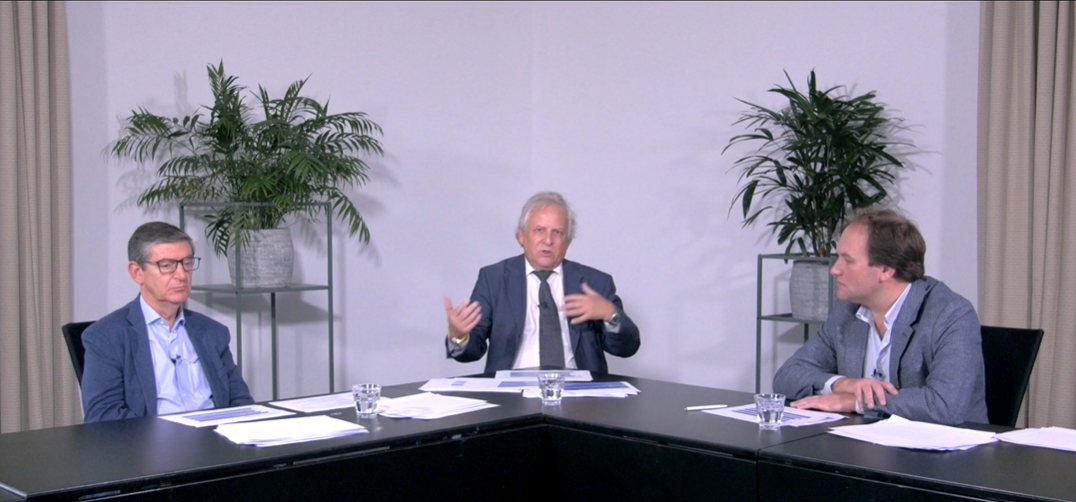
Please find below a link to access the new Oruen Round Table. Please be advised that this is for medical professionals.
This is a peer to peer discussion on the role of anticoagulation in the setting of cerebral venous thromboembolism.
- CVT is a rare type of stroke and peripheral venous thromboembolism
- There is scientific evidence supporting anticoagulation in the acute phase of CVT but scant data to guide longer-term oral anticoagulation
- Non-vitamin-K oral anticoagulants (NOACs) are being investigated as an alternative to warfarin in patients with CVT for longer-term management
This discussion’s audience is for the general neurologists and stroke specialists across Europe, once completed, learners will be able to:
- Discuss risk of venous thromboembolism (VTE) after cerebral venous thrombosis (CVT)
- Explain the design of RE-SPECT CVT trial
- Analyse outcomes in RE-SPECT CVT trial
(please note this content is not available for physicians in the UK & US)
Faculty:
José Ferro
Department of Neurosciences and Mental Health
Hospital Santa Maria/Centro Hospitalar Universitário Lisboa Norte
Lisbon, Portugal
Hans-Christoph Diener
Faculty of Medicine
University Duisburg-Essen
Essen, Germany
Jonathan Coutinho
Department of Neurology
Academic Medical Center University of Amsterdam
Amsterdam, The Netherlands
This round table discussion was filmed in Amsterdam by Oruen and was supported by Boehringer Ingelheim.
This discussion has been awarded 1 CME credit by the EACIC; instructions on how to obtain your CME credit will follow at the end of this video.
Feb 7, 2020
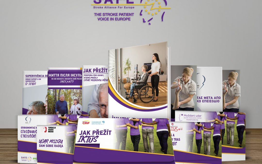
Since 2018, SAFE has participated in the Angels Initiative project providing patient perspective and enabling better patient-doctor communication. The project was so far realised in two phases. In the first phase the patient-centered, evidence-based brochures were translated to 13 languages and distributed in 12 countries, in designated stroke hospitals. In the second phase, conducted in late 2019 and early 2020, the project expanded to include even more countries and languages, increasing reach of these information in 15 European countries.
“It’s proven great to have these books and be able to use them as a tool for raising awareness of stroke prevention and treatment among patients, caregivers and people interested in stroke.”
Hrvoje Budinčević
President of the Croatian Stroke Society / Hrvatsko društvo za prevenciju moždanog udara
The brochures were differently formatted, this time being comprised in one booklet containing five types of stroke-related information. SAFE is proud to report that in the second phase 17 out of 34 its stroke support member organisations participated, making these materials available in 14 languages: Spanish, Serbian, Croatian, Catalan, Georgian, Ukrainian, Greek (Greece), Slovakian, Polish, Turkish, Macedonian, Greek (Cyprus), French, Czech and Hungarian.
“International collaboration of SSOs obviously implies exchange of experience and sharing best examples of materials, ideas, projects, etc. From this point preparing of the set of translated and localized education materials on stroke for patients and general population, initially elaborated by one of the leading SSOs in the world, UK Stroke Association, translated to local languages by national SSOs in several countries with support of SAFE and Angels Initiative, is a great experience, showing all of us that we are not alone, and inspiring us for further own efforts.”
Dmitriy Gulyayev
Director of education, research and publishing projects
Ukrainian Anti-Stroke Association
The project was a great success in all countries where it was presented, clearly demonstrating that there was a gap in availability of stroke-related information among patients. Subsequently, it also brought additional quality to doctor-patient communication in hospitals where patients received these books.
The information provided in the books were kindly provided by the Stroke Association UK*and then translated to all project languages, applying the information standard procedure for the translation.
The original brochures are in English language and you can access their original content by following the links below:
Transient ischaemic attack
Next steps after stroke
Supporting a stroke survivor
When you have a stroke
How to reduce your risk
“The Angels project and the brochures are helping us to connect patient voices. I see in it the potential for a possible change in the healthcare system. It means in cooperation with Ministry of Health to set up guidelines which will contain uniform information, for what are patients after stroke entitled in social, physiotherapy or speech therapy sphere.”
Alžbeta Husarovič, President of Porazka.sk
On this link you can find PDF versions in following languages: Greek (for Greece and Cyprus), Hungarian, Czech, Croatian, Serbian, Macedonian, Turkish, Ukrainian, Georgian, Spanish, Catalan, Hungarian, Polish, Slovakian and French.
SAFE Angels Patient Books for Download
To access the last year’s version of books in Latvian language, please click here.
“SAFE Angels books offer consistency of messages, availability of the required information and ability to share this information. Apart from health-related information, they offer plenty of beneficial advices and solutions for obstacles that patients and their caregivers face every day. After using these books for two years, we think they have helped patients to improve their knowledge, compliance to treatment, self-confidence and self management after stroke.”
Maja Bozinovska Smiceska
President of the Macedonian Stroke Association “Mozočen Udar”
SAFE would like to thank and acknowledge support received from our member stroke support organisations who participated in this project:
Serbia: Serbian Stroke Association
Croatia: Croatian Stroke Society
Spain (Catalonia): Fundacio Ictus
Spain: Federacion Espanola de Ictus
Czech Republic: Sdruzeni SMP
Czech Republic: Cerebrum
Georgia: Medical Foundation Mkurnali
Ukraine: Victory over Stroke
Ukraine: Ukrainian Anti-Stroke Association
Hungary: Stroke Liga Nemzeti
Poland: Fundacja Udaru Mozgu
Macedonia: Macedonian Stroke Association “Mozočen Udar”
Greece: HAAS
Turkey: Beyinder
France: France AVC
Slovakia: Porazka.sk
Cyprus: Cyprus Stroke Association
Luxembourg: Bletz ASBL
About the joint SAFE Angels Initiative project
The mission of Boehringer Ingelheim’s (BI’s) Angels Initiative is to increase the number of patients who can be treated in stroke-ready hospitals and to optimise the quality of treatment in all existing stroke centres. The Stroke Alliance for Europe (SAFE) has partnered with BI to support this ambitious project, adding an important dimension: the patient perspective. SAFE’s involvement means patient-focused information will be available to patients and their carers as soon as they arrive in all stroke units.
About Angels Initiative
Every 30 minutes a stroke patient who could have been saved, dies or is permanently disabled, because they were treated in the wrong hospital.
Angels Initiative is building a global community of stroke centres and stroke-ready hospitals, working every day to improve the quality of treatment for every stroke patient.
In 2017, the Angels Initiative was endorsed by European Stroke Organisation (ESO), the largest European organisation of stroke professionals.
For more information about Angels Initiative, please visit www.angels-initiative.com
Feb 6, 2020
Nenad Nikolić, Stroke Association Serbia’s Secretary
Author: Nenad Nikolić, Stroke Association Serbia’s Secretary and Stroke Survivor’s #BrainLifeGoals project manager
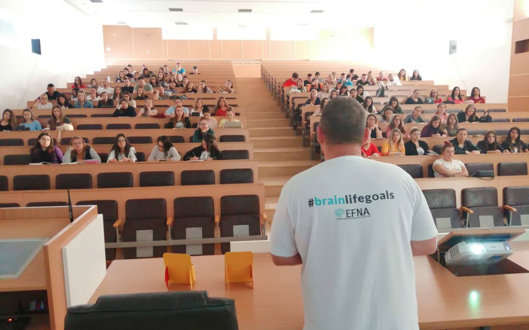
During the Brain awareness week, held 11- 17th March 2019, all around the world, many of actions and campaigns took place, aiming to raise public awareness of the progress and benefits of brain research. It was a chance to inform people about the progress in diagnosing, preventing and treating brain disorders. Our Stroke Association Serbia is involved in European and World stroke campaigns, so we used the opportunity to inform our followers about it, using social media and our website.
It was around that time that the European Federation of Neurological Associations launched #BrainLifeGoals campaign. The goal of this campaign is to show and explore aspirations and desires of people who live with brain disorders. Hashtag #Lifegoals has become a popular trend among social media users, clustering their shares about goals and achievements. Sometimes those goals were to have some clothes designed by a famous fashion designer, to earn a lot of money, to travel to exotic destinations, drive a new car… For a person with a brain disorder, these are not important goals- this is luxury, because these people strive for some „basic“ things such as being able to walk again, to read, to write, to talk etc. Things on a daily basis that most of us do automatically and easily can be an achievement and a life goal for someone with brain disorders. We can call them #BrainLifeGoals.
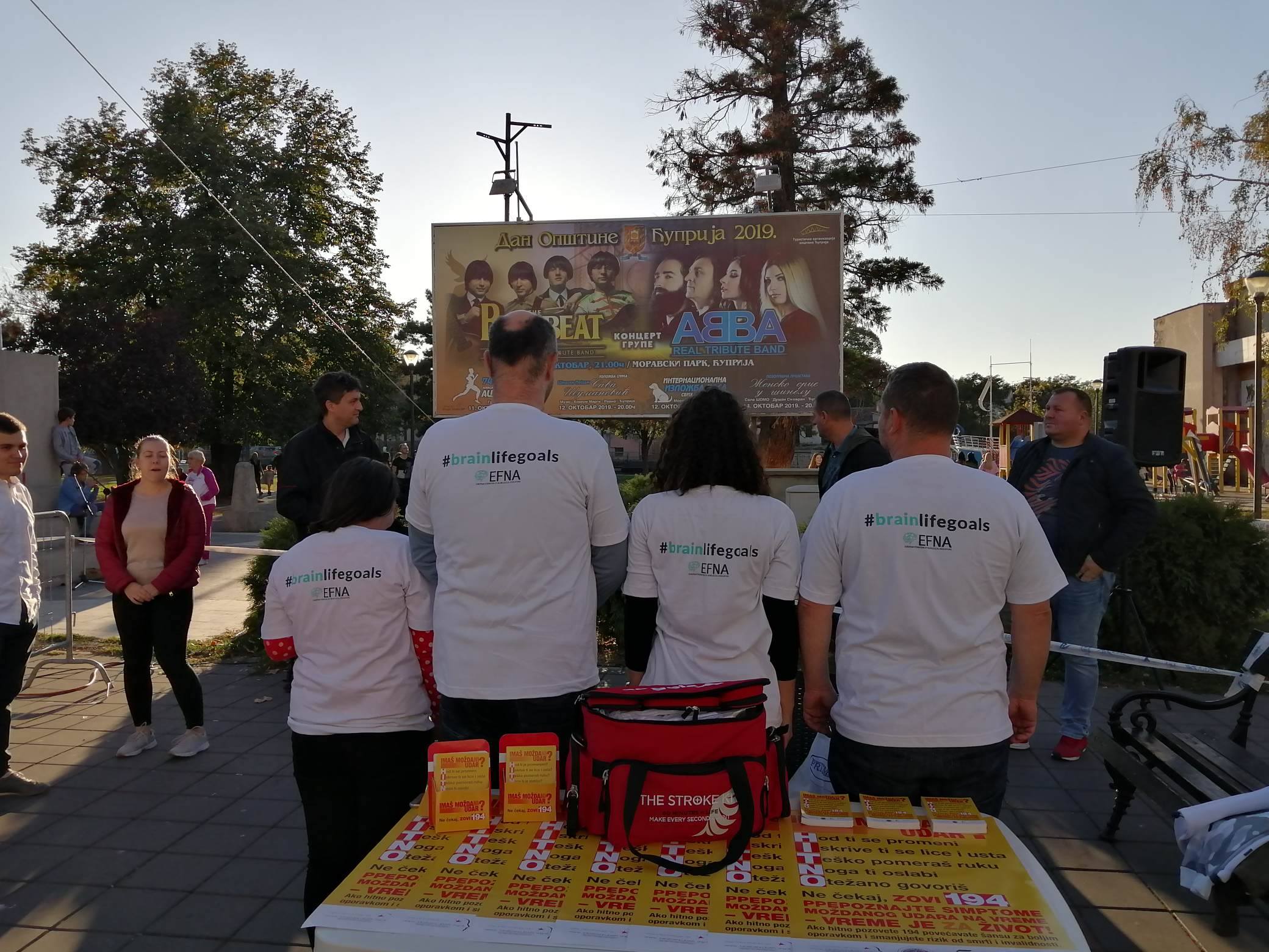 Our Association recognized the importance of this particular campaign because we could easily relate to it. We decided to support it with active participation. Since I have been managing our Association’s Facebook and Twitter page, and have a very active communication with our followers, I was privileged to receive many people’s stories about their personal experience with stroke.
We decided to publish stories of people with stroke, with an emphasis on their #BrainLifeGoals during their recovery. Than we asked and encouraged those people, most of being in their twenties and early thirties, to share their stories publicly, and to raise awareness of all the problems they’re facing, but also to encourage others with stroke and give them strength to continue their daily struggle with stroke consequences.
No matter how hard we try to advocate for stroke, no one can do it better than someone who has experienced it.
Preparing these stories was a bit of challenge, because there is a lot to be said, and I needed to prioritize. I felt a lot of responsibility, because I was writing about someone’s life, and it had to be done the right way and without many medical terms in order to make understandable for broader audience. The facts and events they told us were shaping stories. Each story was different and had its own concept. All these stories contain variety of emotions: Fear, anxiety, uncertainty, but also hope and huge amount of willpower and support. These stories carry strong messages. Almost every storyteller’s life has changed drastically and these people have now completely different views on life and its aspects, many of them have new hobbies, new healthy habits, many changed their nutrition habits to healthy diets, many of them became more stress tolerant… Highlighting these changes felt like a very important thing to do.
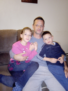
Predrag M. – A middle school physics teacher, a father and a stroke survivor whose #BrainLifeGoal was to be able to read again.
Their #BrainLifeGoals vary from person to person. For someone it was babysitting and playing with their children, for someone cycling, reading, walking without help to nearby sightseeing… Eight stories have been published so far, and some of storytellers had a stroke at the age of 18 and 19! Several more stories are in preparation and will be published soon. Some of our story tellers- Stroke Survivors, had a stroke in their sleep, some while on work. It is clear that stroke can happen anytime, anywhere, to anyone. All these stories have a strong empathetic potential and when read, the reader is faced with situations and problems that the survivors are facing every day. If these stories are encouraging people to think that way, it is our victory! We modified our website www.mozdaniudar.org so every single published story is on one page that can be easily accessed with only one click www.mozdaniudar.org/wp/brainlifegoals/
Except for the important messages stroke survivors tell, it is very important to underline that they also talk openly about stroke. This is, once again, very important, because these wonderful people are encouraging others with stroke to talk freely about their problems, and prevent stigmatization of stroke patients.
The usual procedure is that we first share story on our website and social media accounts, than we translate it to English, and later EFNA shares them on their site www.efna.net/brainlifegoals/ . This way, stories of stroke survivors reach larger international audience. Other Internet portals sometimes share our stories, and we are happy about it. We started with a story of a mother of three who suffered a stroke at 40. Her basic motivation and #BrainLifeGoal was to play again with her children. We continued Campaign with a story of young nursery teacher who had a stroke, and after she recovered, she won a medal in downhill cycling! This inspiring story shows that not only recovery is possible, but also excellent results can be achieved.

Marina K. – A Downhill Biking Champion, a mother and a stroke survivor
In all our cover stories focus is on a stroke survivor, which is very important. For me personally, participating and managing Campaign #BrainLifeGoals in Serbia is a big pleasure, and I really enjoy working on this. I am very proud that our participating in this campaign with working title “Stroke survivor’s brain life goals” is rewarded by EFNA with a grant that really meant a lot to our organisation and helped us organize our core activities.
We are all excited because we feel that we are doing a right thing, helping to raise public awareness about stroke and difficulties that stroke survivors are experiencing in their life after stroke. In a way, we became a “PATIENT VOICE” which is one of our SSO’s basic purposes, and a slogan of the Stroke Alliance for Europe whose member we are proud to be – “The Stroke Patient Voice in Europe”.
Image credits: All images used in this article are property of the Stroke Association Serbia.
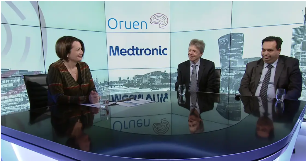
Feb 4, 2020
Professor Valeria Caso, Associate Professor of Neurology, University of Perugia Stroke Unit, Perugia, Italy, Professor Helmut Pürerfellner, Department of Cardiology, Public Hospital Elisabethinen, Linz, Austria and Professor Georgios Tsivgoulis, Associate Professor of Neurology, Second Department of Neurology, National & Kapodistrian University of Athens, Greece discussed the different types of stroke and how it is a major healthcare issue. They also discussed the value of prolonged monitoring in selected ESUS patients, and also some new data that might reconfirm what we already know – that if we have longer monitoring in some of those patients, then we will have even better outcomes.
Learning Objectives
- New evidence and increasing the awareness of atrial fibrillation (AF) in cryptogenic stroke patients.
- Treatment approaches from the guidelines and the latest study evidence.
- AF, stroke, and related symptoms, and why the evidence from the past is important.
- Why ESUS needs prolonged monitoring and how new data may benefit patients.
For accessing the webinar video, please click on the button below:
Oruen Webinar
For further information please visit Oruen website.
This webinar was supported by Medtronic.
Jan 31, 2020
First published on ScenceDaily.com
New research from the Smidt Heart Institute at Cedars-Sinai showed for the first time that women’s blood vessels — including both large and small arteries — age at a faster rate than men’s. The findings, published Wednesday in the journal JAMA Cardiology, could help to explain why women tend to develop different types of cardiovascular disease and with different timing than men.
“Many of us in medicine have long believed that women simply ‘catch up’ to men in terms of their cardiovascular risk,” said Susan Cheng, MD, MPH, MMSc, senior author of the study and director of Public Health Research at the Smidt Heart Institute. “Our research not only confirms that women have different biology and physiology than their male counterparts, but also illustrates why it is that women may be more susceptible to developing certain types of cardiovascular disease and at different points in life.”
Using community-based data amassed from multiple sites across the country, Cheng and her research team conducted sex-specific analyses of measured blood pressure — a critical indicator of cardiovascular risk. The data represented nearly 145,000 blood pressure measurements, collected serially over a 43-year period, from 32,833 study participants ranging in age from 5 to 98 years old.
Because a person’s risk for developing a heart attack, heart failure, or a stroke typically begins with having high blood pressure, Cedars-Sinai researchers combed through their massive data looking for clues and patterns regarding how blood pressure starts to rise. Then, instead of comparing the data from men and women to each other, investigators compared women to women and men to men.
This approach allowed investigators to identify that the progression and evolution of women’s vascular function is very different than for men. In fact, women showed signs of blood pressure elevation much earlier in life than men.
“Our data showed that rates of accelerating blood pressure elevation were significantly higher in women than men, starting earlier in life,” said Cheng, the Erika J. Glazer Chair in Women’s Cardiovascular Health, who also serves as director of Cardiovascular Population Sciences at the Barbra Streisand Women’s Heart Center. “This means that if we define the hypertension threshold the exact same way, a 30-year old woman with high blood pressure is probably at higher risk for cardiovascular disease than a man with high blood pressure at the same age.”
Christine Albert, MD, MPH, founding chair of the newly established Department of Cardiology at the Smidt Heart Institute, says this new research should help guide clinicians and researchers to think differently when it comes to treating and studying women and their cardiovascular health.
You can read the full article here.
Image: Pixabay