Aug 29, 2018
Published on ScienceDaily.com
Four out of ten patients with atrial fibrillation but no history of stroke or transient ischaemic attack have previously unknown brain damage, according to the first results of the Swiss Atrial Fibrillation Cohort Study (Swiss-AF) presented today at ESC Congress 2018.
“Our results suggest that clinically unrecognised brain damage may explain the association between dementia and atrial fibrillation in patients without prior stroke,” said Co-Principal Investigator Professor David Conen of McMaster University, Hamilton, Canada.
Patients with atrial fibrillation have a significantly increased risk of stroke, which is why most are treated with blood thinners (oral anticoagulation). This increased stroke risk is probably the main reason why patients with atrial fibrillation also face an increased risk of cognitive dysfunction and dementia. However, the relationship between atrial fibrillation and dementia has also been shown among patients without prior strokes, meaning that additional mechanisms have to be involved.
Clarifying the mechanisms by which atrial fibrillation increases the risk of cognitive dysfunction and dementia is a first step towards developing preventive measures.
Swiss-AF is a prospective, observational study designed to pinpoint the mechanisms of cognitive decline in patients with atrial fibrillation.2 This analysis investigated the prevalence of silent brain damage in atrial fibrillation patients.
The study enrolled 2,415 patients aged over 65 years with atrial fibrillation between 2014 and 2017 from 14 centres in Switzerland. All patients without contraindications underwent standardised brain magnetic resonance imaging and the images were analysed in a central core laboratory. Scans were available in 1,736 patients. Of those, 347 (20%) patients had a history of stroke and/or transient ischaemic attack and were excluded from the analysis.
The final analysis included 1,389 patients with atrial fibrillation but no history of stroke or transient ischaemic attack. The average age of participants was 72 years, and 26% were women. The scans showed that 569 (41%) patients had at least one type of previously unknown brain damage: 207 (15%) had a cerebral infarct, 269 (19%) had small bleeds in the brain (microbleeds), and 222 (16%) had small deep brain lesions called lacunes.
“Four in ten patients with atrial fibrillation but no history of stroke or transient ischaemic attack had clinically unrecognised ‘silent’ brain lesions,” said Professor Conen. “This brain damage could trigger cognitive decline.”
Most study participants (1,234; 89%) were treated with oral anticoagulants. Co-Principal investigator Professor Stefan Osswald of University Hospital Basel, Switzerland, noted that the cross-sectional analysis looked at the data at a single point in time and cannot address the question of whether the cerebral infarcts and other brain lesions occurred before or after initiation of oral anticoagulation. But he said: “The findings nevertheless raise the issue that oral anticoagulation might not prevent all brain damage in patients with atrial fibrillation.”
Professor Conen said: “All Swiss-AF participants underwent extensive cognitive testing. These data will be analysed to see whether patients with silent brain lesions also have impaired cognitive function.” Collaborations with other study groups will help to sort out whether these findings are specific to patients with atrial fibrillation.
Story Source:”Four out of 10 patients with atrial fibrillation have unknown brain damage.” ScienceDaily. ScienceDaily, 26 August 2018. <www.sciencedaily.com/releases/2018/08/180826120744.htm>.

Aug 24, 2018
The following content was first published on EU Commission official website
According to a recently published study, European patients are still generally unaware of their rights and the possibility to access health services in other EU Member States, as well as of the existence of National Contact Points (NCPs). But the situation is improving.
National Contact Points (NCPs) aim to help patients exercise their rights under the Cross-border Healthcare Directive. But how can they improve their work?
Using a combination of research methods, including a literature review, an analysis of legal texts, a website analysis, a pseudo-patient investigation, and surveys of NCPs and patients, the aim of the study carried out by Ecorys, KU Leuven and GfK Belgium was to identify how to improve the current level of information on cross-border healthcare available to patients.
Websites
The study found that although the information available to patients on NCP websites was adequate, the websites themselves need improvements, especially the sections on patients’ rights (for incoming patients), quality and safety standards (for incoming patients) and reimbursement of cross-border healthcare costs (for outgoing patients).
However, compared to the results of the earlier Evaluative study(fieldwork carried out in 2014), the NCPs have made significant progress in this area.
Toolbox and training material
This study has also resulted in the development of a practice-orientated toolbox and training material to help the NCPs improve the quality of information for patients, as well as a set of Guiding Principles and indicators for establishing an NCP service that is more uniform, patient-centred and in line with the legal requirements. This will contribute to high level information provision to patients.
The study feeds into the upcoming implementation report on the operation of the Cross-border Healthcare Directive due this October.
More information
Aug 6, 2018
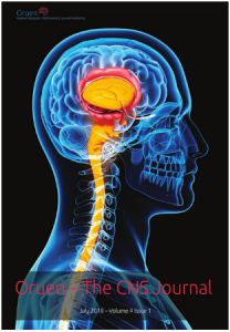
Oruen – The CNS Journal is a peer-reviewed, open access publication, and has received CME accreditation from the European Accreditation Committee in CNS (EACIC), with a 100% focus on original CNS research topics, and the latest advances, diagnoses, and treatment of CNS disorders.
The Journal is distributed in print and electronically to thousands of physicians, researchers, academics, nurses, and related healthcare professionals with an interest in CNS disorders. Both subscription and access are free and there are no contributory author fees for publication. Papers submitted for publication are accepted based on their originality, likely impact on and relevance to clinical practice, data quality, and overall potential interest to the journal’s readership.
Oruen – The CNS Journal is published bi-annually. The first issue of the journal was published in May 2015
You can access the latest issue by clicking on the photo below:
For any questions or submission requests/enquiries please contact Dr James Coe – Head Editor editor@oruen.com
Jul 27, 2018
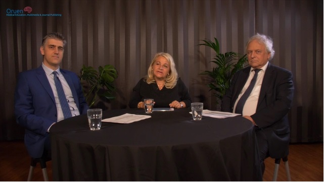
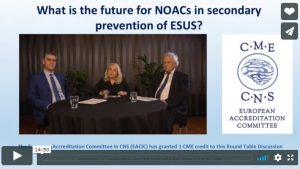
The 15 min round table discussion by Christina Sjöstrand, Stockholm, Hans-Christoph Diener, Essen and George Ntaios, Thessaly, focuses on the concepts of randomized clinical trials for secondary stroke prevention in patients with embolic stroke of undetermined source (ESUS). Two large clinical trials tested the hypothesis that non-vitamin K antagonist oral anticoagulants (NOACs) might be superior to aspirin in preventing recurrent strokes after ESUS.
ESUS is a type of ischemic stroke with unknown origin, i.e. for which no probable cause can be identified after standard diagnostic evaluation. Thus, ESUS is a subgroup of cryptogenic stroke, which also includes strokes with incomplete evaluation and those for which more than one probable cause. Non-lacunar strokes in patients without no major-risk cardioembolic source (such as atrial fibrillation), no major atherosclerosis of the arteries supplying the brain infarct area and no other specific cause of their stroke are identified as ESUS.
Current strategy for secondary stroke prevention is based on antiplatelets but stroke recurrent rates remain high. A historical trial, WARSS, pointed at a potential advantage of anticoagulation over antiplatelet in patients with cryptogenic stroke. There is broad evidence for anticoagulation for stroke prevention in atrial fibrillation, thus providing another rationale for testing anticoagulation in ESUS.
Two large trials randomized ESUS patients for long-term treatment with either a NOAC or aspirin: NAVIGATE ESUS was stopped after an interim analysis as efficacy was not different with rivaroxaban compared with aspirin but showed an increased risk of major bleeding. RE-SPECT ESUS is still ongoing as planned; in this trial dabigatran is tested against aspirin. Results will be presented in October 2018. If the trial is positive, i.e. showing greater efficacy of dabigatran in the prevention of stroke recurrence than aspirin, this may not only have great implications of how ESUS patients are treated but could also lead to a simplified post-stroke diagnostic workup for most patients with ischemic stroke.
Please click on the photo below to access the round table video.

Jul 17, 2018
Having a stroke is a terrifying experience, and it is only natural to start experiencing some symptoms of anxiety and depression when it is all over. After all, it is something that often brings a person close to death, and it can seem like no one else understands the feelings of fear and despair that often accompany survival. While you often feel thankful that you were able to make it through, there is nothing wrong with the negative feelings that come with it. Here is how your canine companion can help you get through this tough phase after a stroke.
Alleviating Anxiety and Depression
These are natural things to feel after a stroke, but a dog can help you get through them. Petting and hugging them releases oxytocin throughout the body, and this is the hormone responsible for reducing anxiety and stress, blood pressure, and heart rate, so you feel more relaxed and a lot less worried. They also bring comfort as a whole because you know they are always there when you need them.
They give you a sense of purpose and validation, as well as offering unconditional love that cannot be matched by another creature on this planet. There have even been studies to suggest that they are able to help the process of balancing the serotonin levels in your brain, boosting your mood and leaving you feeling in a better mindset.
Reducing Feelings of Loneliness
Often, post-stroke life can leave a person feeling increasingly lonely, and this can happen for a number of reasons. Sometimes, a lot of attention will have been received in the hospital, and family will have made special arrangements to come visit and be there as much as possible.
However, once everything is clear, life goes back to normal, and it can leave a person feeling isolated. A dog is always there to offer love, support, and companionship, and this can bring a great deal of comfort, as well as reduce feelings of loneliness and isolation.
Giving You Someone to Talk to
Possibly the most important thing that a dog can do for you is give you someone to talk to when you need it most. They will not judge or abandon you, regardless of how emotional you are. A shoulder to cry on, there is nothing you cannot tell them – especially as they will never tell anyone else. Linking to the fact that they can help with loneliness, it is important to have someone to speak to in these circumstances, and sometimes another human is not the easiest or best choice for you. A dog can offer just as much, and more.
To Conclude
If you found this interesting and would like to learn more about the ways in which a dog can help with mental health, feel free to check out this detailed resource. Dogs are some of the greatest companions we could ever ask for, and as little as we give them, they always return it tenfold. We understand the struggle that comes after a stroke, and your dog is there to help you through the darkest moments in every way they can.
About the Author

Will Tottle
Will Tottle is a freelance writer, his blog can be seen here . If you are interested in more information on the benefits of dog ownership including health tips, buyer’s guides and gear reviews, then check out his guides over at Dogowner.co.uk
Follow on Facebook or Twitter

Jun 24, 2018
First appeared on ScienceDaily.com
Paralysis of an arm and/or leg is one of the most common effects of a stroke. But thanks to research carried out by scientists at the Defitech Foundation Chair in Brain-Machine Interface, in association with other members of EPFL’s Center for Neuroprothetics, the Clinique Romande de Réadaptation in Sion, and the Geneva University Hospitals, stroke victims may soon be able to recover greater use of their paralyzed limbs. The scientists’ pioneering approach brings together two known types of therapies — a brain-computer interface (BCI) and functional electrical stimulation (FES) — and has been published in Nature Communications.
“The key is to stimulate the nerves of the paralyzed arm precisely when the stroke-affected part of the brain activates to move the limb, even if the patient can’t actually carry out the movement. That helps reestablish the link between the two nerve pathways where the signal comes in and goes out,” says José del R. Millán, who holds the Defitech Chair at EPFL.
Twenty-seven patients aged 36 to 76 took part in the clinical trial. All had a similar lesion that resulted in moderate to severe arm paralysis following a stroke occurring at least ten months earlier. Half of the patients were treated with the scientists’ dual-therapy approach and reported clinically significant improvements. The other half were treated only with FES and served as a control group.
For the first group, the scientists used a BCI system to link the patients’ brains to computers using electrodes. That let the scientists pinpoint exactly where the electrical activity occurred in the brain tissue when the patients tried to reach out their hands. Every time that the electrical activity was identified, the system immediately stimulated the arm muscle controlling the corresponding wrist and finger movements. The patients in the second group also had their arm muscles stimulated, but at random times. This control group enabled the scientists to determine how much of the additional motor-function improvement could be attributed to the BCI system.
Reactivated tissue
The scientists noted a significant improvement in arm mobility among patients in the first group after just ten one-hour sessions. When the full round of treatment was completed, some of the first-group patients’ scores on the Fugl-Meyer Assessment — a test used to evaluate motor recovery among patients with post-stroke hemiplegia — were over twice as high as those of the second group.
“Patients who received the BCI treatment showed more activity in the neural tissue surrounding the affected area. Due to their plasticity, they could help make up for the functioning of the damaged tissue,” says Millán.
Electroencephalographies (EEGs) of the patients clearly showed an increase in the number of connections among the motor cortex regions of their damaged brain hemisphere, which corresponded with the increased ease in carrying out the associated movements. What’s more, the enhanced motor function didn’t seem to diminish with time. Evaluated again 6-12 months later, the patients hadn’t lost any of their recovered mobility.
Story Source: Ecole Polytechnique Fédérale de Lausanne. “A dual-therapy approach to boost motor recovery after a stroke.” ScienceDaily. ScienceDaily, 20 June 2018. <www.sciencedaily.com/releases/2018/06/180620094808.htm>.