Sep 3, 2018
The original article published at ScienceDaily.com
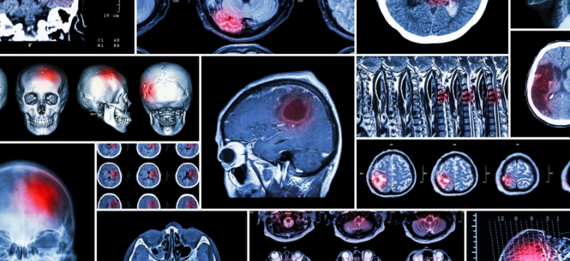
People who have had a stroke are around twice as likely to develop dementia, according to the largest study of its kind ever conducted.
The University of Exeter Medical School led the study which analysed data on stroke and dementia risk from 3.2 million people across the world. The link between stroke and dementia persisted even after taking into account other dementia risk factors such as blood pressure, diabetes and cardiovascular disease. Their findings give the strongest evidence to date that having a stroke significantly increases the risk of dementia.
The study builds on previous research which had established the link between stroke and dementia, though had not quantified the degree to which stroke actually increased dementia risk. To better understand the link between the two, researchers analysed 36 studies where participants had a history of stroke, totalling data from 1.9 million people. In addition, they analysed a further 12 studies that looked at whether participants had a recent stroke over the study period, adding a further 1.3 million people. The new research, published in the leading dementia journal Alzheimer’s & Dementia: The Journal of the Alzheimer’s Association, is the first meta-analysis in the area.
Dr Ilianna Lourida, of the University of Exeter Medical School, said: “We found that a history of stroke increases dementia risk by around 70%, and recent strokes more than doubled the risk. Given how common both stroke and dementia are, this strong link is an important finding. Improvements in stroke prevention and post-stroke care may therefore play a key role in dementia prevention.”
According to the World Health Organisation, 15 million people have a stroke each year. Meanwhile, around 50 million people globally have dementia – a number expected to almost double ever 20 years, reaching 131 million by 2050.
Stroke characteristics such as the location and extent of brain damage may help to explain variation in dementia risk observed between studies, and there was some suggestion that dementia risk may be higher for men following stroke.
Further research is required to clarify whether factors such as ethnicity and education modify dementia risk following stroke. Most people who have a stroke do not go on to develop dementia, so further research is also needed to establish whether differences in post-stroke care and lifestyle can reduce the risk of dementia further.
Dr David Llewellyn, from the University of Exeter Medical School, concluded: “Around a third of dementia cases are thought to be potentially preventable, though this estimate does not take into account the risk associated with stroke. Our findings indicate that this figure could be even higher, and reinforce the importance of protecting the blood supply to the brain when attempting to reduce the global burden of dementia.”
Story Source: University of Exeter. “Stroke doubles dementia risk, concludes large-scale study.” ScienceDaily. ScienceDaily, 31 August 2018. <www.sciencedaily.com/releases/2018/08/180831083542.htm>.

Aug 29, 2018
Published on ScienceDaily.com
Postmenopausal factors may have an impact on the heart-protective qualities of high-density lipoproteins (HDL) — also known as ‘good cholesterol’ — according to a study led by researchers in the University of Pittsburgh Graduate School of Public Health.
The findings, published today in Arteriosclerosis, Thrombosis, and Vascular Biology, a journal of the American Heart Association (AHA), indicate that this specific type of blood cholesterol may not translate into a lowered risk of cardiovascular disease in older women — bringing into question the current use of HDL cholesterol in a common equation designed to predict heart disease risk, particularly for women.
HDL is a family of particles found in the blood that vary in sizes and cholesterol contents. HDL has traditionally been measured as the total cholesterol carried by the HDL particles, known as HDL cholesterol. HDL cholesterol, however, does not necessarily reflect the overall concentration, the uneven distribution, or the content and function of HDL particles. Previous research has demonstrated the heart-protective features of HDL. This good cholesterol carries fats away from the heart, reducing the build-up of plaque and lowering the potential for cardiovascular disease.
“The results of our study are particularly interesting to both the public and clinicians because total HDL cholesterol is still used to predict cardiovascular disease risk,” said lead author Samar R. El Khoudary, Ph.D., M.P.H., F.A.H.A., associate professor in Pitt Public Health’s Department of Epidemiology. “This study confirms our previous work on a different group of women and suggests that clinicians need to take a closer look at the type of HDL in middle-aged and older women, because higher HDL cholesterol may not always be as protective in postmenopausal women as we once thought. High total HDL cholesterol in postmenopausal women could mask a significant heart disease risk that we still need to understand.”
El Khoudary’s team looked at 1,138 women aged 45 through 84 enrolled across the U.S. in the Multi-Ethnic Study of Atherosclerosis (MESA), a medical research study sponsored by the National Heart, Lung and Blood Institute of the National Institutes of Health (NIH). MESA began in 1999 and is still following participants today.
The study points out that the traditional measure of the good cholesterol, HDL cholesterol, fails to portray an accurate depiction of heart disease risk for postmenopausal women.
Women are subject to a variety of physiological changes in their sex hormones, lipids, body fat deposition and vascular health as they transition through menopause. The authors are hypothesizing that the decrease of estrogen, a cardio-protective sex hormone, along with other metabolic changes, can trigger chronic inflammation over time, which may alter the quality of HDL particles.
“We have been seeing an unexpected relationship between HDL cholesterol and postmenopausal women in previous studies, but have never deeply explored it,” said El Khoudary. Her study looked at two specific measurements of HDL to draw the conclusion that HDL cholesterol is not always cardio-protective for postmenopausal women, or not as ‘good’ as expected.
The number and size of the HDL particles and total cholesterol carried by HDL particles was observed. The study also looked at how age when women transitioned into postmenopause, and the amount of time since transitioning, may impact the expected cardio-protective associations of HDL measures.
The harmful association of higher HDL cholesterol with atherosclerosis risk was most evident in women with older age at menopause and who were greater than, or equal to, 10 years into postmenopause.
In contrast to HDL cholesterol, a higher concentration of total HDL particles was associated with lower risk of atherosclerosis. Additionally, having a high number of small HDL particles was found beneficial for postmenopausal women. These findings persist irrespective of age and how long it has been since women became postmenopausal.
On the other hand, large HDL particles are linked to an increased risk of cardiovascular disease close to menopause. During this time, the quality of HDL may be reduced, increasing the chance for women to develop atherosclerosis or cardiovascular disease. As women move further away from their transition, the quality of the HDL may restore — making the good cholesterol cardio-protective once again.
“Identifying the proper method to measure active ‘good’ HDL is critical to understanding the true cardiovascular health of these women,” said senior author Matthew Budoff, M.D., of Los Angeles Biomedical Research Institute.
El Khoudary recently was awarded funding from the National Institute on Aging to expand upon this research work. Her goal is to continue understanding the link between quality of good cholesterol over the menopause transition and women’s risk of cardiovascular disease later in life. She also seeks to examine the biological mechanisms that contribute to quality change of good cholesterol, so that the cardio-protective contribution of good cholesterol to postmenopausal women’s health can be clarified, which would impact guidelines for screening and treatment.
Additional authors on this study are Indre Ceponiene, M.D., Ph.D., of Harbor-UCLA Medical Center and Lithuanian University of Health Sciences; Saad Samargandy, M.P.H., of Pitt; James H. Stein, M.D., and Matthew C. Tattersall, D.O., M.S., both of University of Wisconsin; Dong Li, Ph.D., of Los Angeles Biomedical Research Center in Torrance CA.
This research was funded by NIH grants R01 HL071739, N01-HC-95159, N01-HC-95160, N01-HC-95161, N01-HC-95162, N01-HC-95163, N01-HC-95164, N01-HC-95165, N01-HC-95166, N01-HC-95167, N01-HC-95168, N01-HC-95169, UL1-TR-000040, UL1 TR 001079 and UL1-RR-025005; and a grant from Quest Diagnostics.
Story Source: Chicago, University of Pittsburgh Schools of the Health Sciences. “‘Good cholesterol’ may not always be good.” ScienceDaily. ScienceDaily, 19 July 2018. <www.sciencedaily.com/releases/2018/07/180719085420.htm>.

Aug 29, 2018
Published on ScienceDaily.com
Four out of ten patients with atrial fibrillation but no history of stroke or transient ischaemic attack have previously unknown brain damage, according to the first results of the Swiss Atrial Fibrillation Cohort Study (Swiss-AF) presented today at ESC Congress 2018.
“Our results suggest that clinically unrecognised brain damage may explain the association between dementia and atrial fibrillation in patients without prior stroke,” said Co-Principal Investigator Professor David Conen of McMaster University, Hamilton, Canada.
Patients with atrial fibrillation have a significantly increased risk of stroke, which is why most are treated with blood thinners (oral anticoagulation). This increased stroke risk is probably the main reason why patients with atrial fibrillation also face an increased risk of cognitive dysfunction and dementia. However, the relationship between atrial fibrillation and dementia has also been shown among patients without prior strokes, meaning that additional mechanisms have to be involved.
Clarifying the mechanisms by which atrial fibrillation increases the risk of cognitive dysfunction and dementia is a first step towards developing preventive measures.
Swiss-AF is a prospective, observational study designed to pinpoint the mechanisms of cognitive decline in patients with atrial fibrillation.2 This analysis investigated the prevalence of silent brain damage in atrial fibrillation patients.
The study enrolled 2,415 patients aged over 65 years with atrial fibrillation between 2014 and 2017 from 14 centres in Switzerland. All patients without contraindications underwent standardised brain magnetic resonance imaging and the images were analysed in a central core laboratory. Scans were available in 1,736 patients. Of those, 347 (20%) patients had a history of stroke and/or transient ischaemic attack and were excluded from the analysis.
The final analysis included 1,389 patients with atrial fibrillation but no history of stroke or transient ischaemic attack. The average age of participants was 72 years, and 26% were women. The scans showed that 569 (41%) patients had at least one type of previously unknown brain damage: 207 (15%) had a cerebral infarct, 269 (19%) had small bleeds in the brain (microbleeds), and 222 (16%) had small deep brain lesions called lacunes.
“Four in ten patients with atrial fibrillation but no history of stroke or transient ischaemic attack had clinically unrecognised ‘silent’ brain lesions,” said Professor Conen. “This brain damage could trigger cognitive decline.”
Most study participants (1,234; 89%) were treated with oral anticoagulants. Co-Principal investigator Professor Stefan Osswald of University Hospital Basel, Switzerland, noted that the cross-sectional analysis looked at the data at a single point in time and cannot address the question of whether the cerebral infarcts and other brain lesions occurred before or after initiation of oral anticoagulation. But he said: “The findings nevertheless raise the issue that oral anticoagulation might not prevent all brain damage in patients with atrial fibrillation.”
Professor Conen said: “All Swiss-AF participants underwent extensive cognitive testing. These data will be analysed to see whether patients with silent brain lesions also have impaired cognitive function.” Collaborations with other study groups will help to sort out whether these findings are specific to patients with atrial fibrillation.
Story Source:”Four out of 10 patients with atrial fibrillation have unknown brain damage.” ScienceDaily. ScienceDaily, 26 August 2018. <www.sciencedaily.com/releases/2018/08/180826120744.htm>.

Aug 24, 2018
The following content was first published on EU Commission official website
According to a recently published study, European patients are still generally unaware of their rights and the possibility to access health services in other EU Member States, as well as of the existence of National Contact Points (NCPs). But the situation is improving.
National Contact Points (NCPs) aim to help patients exercise their rights under the Cross-border Healthcare Directive. But how can they improve their work?
Using a combination of research methods, including a literature review, an analysis of legal texts, a website analysis, a pseudo-patient investigation, and surveys of NCPs and patients, the aim of the study carried out by Ecorys, KU Leuven and GfK Belgium was to identify how to improve the current level of information on cross-border healthcare available to patients.
Websites
The study found that although the information available to patients on NCP websites was adequate, the websites themselves need improvements, especially the sections on patients’ rights (for incoming patients), quality and safety standards (for incoming patients) and reimbursement of cross-border healthcare costs (for outgoing patients).
However, compared to the results of the earlier Evaluative study(fieldwork carried out in 2014), the NCPs have made significant progress in this area.
Toolbox and training material
This study has also resulted in the development of a practice-orientated toolbox and training material to help the NCPs improve the quality of information for patients, as well as a set of Guiding Principles and indicators for establishing an NCP service that is more uniform, patient-centred and in line with the legal requirements. This will contribute to high level information provision to patients.
The study feeds into the upcoming implementation report on the operation of the Cross-border Healthcare Directive due this October.
More information
Aug 6, 2018
Oruen – The CNS Journal is a peer-reviewed, open access publication, and has received CME accreditation from the European Accreditation Committee in CNS (EACIC), with a 100% focus on original CNS research topics, and the latest advances, diagnoses, and treatment of CNS disorders.
The Journal is distributed in print and electronically to thousands of physicians, researchers, academics, nurses, and related healthcare professionals with an interest in CNS disorders. Both subscription and access are free and there are no contributory author fees for publication. Papers submitted for publication are accepted based on their originality, likely impact on and relevance to clinical practice, data quality, and overall potential interest to the journal’s readership.
Oruen – The CNS Journal is published bi-annually. The first issue of the journal was published in May 2015
You can access the latest issue by clicking on the photo below:
For any questions or submission requests/enquiries please contact Dr James Coe – Head Editor editor@oruen.com

Jul 27, 2018
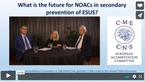
The 15 min round table discussion by Christina Sjöstrand, Stockholm, Hans-Christoph Diener, Essen and George Ntaios, Thessaly, focuses on the concepts of randomized clinical trials for secondary stroke prevention in patients with embolic stroke of undetermined source (ESUS). Two large clinical trials tested the hypothesis that non-vitamin K antagonist oral anticoagulants (NOACs) might be superior to aspirin in preventing recurrent strokes after ESUS.
ESUS is a type of ischemic stroke with unknown origin, i.e. for which no probable cause can be identified after standard diagnostic evaluation. Thus, ESUS is a subgroup of cryptogenic stroke, which also includes strokes with incomplete evaluation and those for which more than one probable cause. Non-lacunar strokes in patients without no major-risk cardioembolic source (such as atrial fibrillation), no major atherosclerosis of the arteries supplying the brain infarct area and no other specific cause of their stroke are identified as ESUS.
Current strategy for secondary stroke prevention is based on antiplatelets but stroke recurrent rates remain high. A historical trial, WARSS, pointed at a potential advantage of anticoagulation over antiplatelet in patients with cryptogenic stroke. There is broad evidence for anticoagulation for stroke prevention in atrial fibrillation, thus providing another rationale for testing anticoagulation in ESUS.
Two large trials randomized ESUS patients for long-term treatment with either a NOAC or aspirin: NAVIGATE ESUS was stopped after an interim analysis as efficacy was not different with rivaroxaban compared with aspirin but showed an increased risk of major bleeding. RE-SPECT ESUS is still ongoing as planned; in this trial dabigatran is tested against aspirin. Results will be presented in October 2018. If the trial is positive, i.e. showing greater efficacy of dabigatran in the prevention of stroke recurrence than aspirin, this may not only have great implications of how ESUS patients are treated but could also lead to a simplified post-stroke diagnostic workup for most patients with ischemic stroke.
Please click on the photo below to access the round table video.
