Nov 3, 2018
Published first on ScienceDaily.com
About 6 million Australians aged 18 years and over have high blood pressure. Of these, more than two thirds had uncontrolled or unmanaged high blood pressure (not taking medication), representing 4 million adult Australians.
High blood pressure, or hypertension, is suggested to be one of the leading risk factors for heart disease.
The process in which high blood pressure causes heart disease is not completely understood.
But now scientists at the Baker Heart and Diabetes Institute have found that high blood pressure caused by specific signalling from the brain promotes heart disease by altering stem cells with the bone marrow.
The results, published in Haematologica demonstrate how an overactive sympathetic nervous system that causes elevated blood pressure can instruct bone marrow stem cells to produce more white blood cells that clog up blood vessels.
The Baker Institute’s Head of Haematopoiesis and Leukocyte Biology, Associate Professor Andrew Murphy says the findings represent a new era of heart disease research.
“Hypertension is a major, independent risk factor of atherosclerotic cardiovascular disease, but we need more information to determine how it is resulting in heart attacks and strokes,” said Associate Professor Murphy.
Atherosclerotic cardiovascular disease is a build-up of cholesterol plaque in the walls of arteries, causing obstruction of blood flow.
“We now know that significance changes in the immune system contributes significantly to heart disease,” he said. “We aimed to determine how the sympathetic nervous system through the brain directly promotes atherosclerosis in the setting of hypertension.”
“We have discovered that this form of high blood pressure, often associated with stress, causes changes within the bone marrow leading to increased white blood cells circulating though our vessels. This is significant as the general view of hypertension is that it is mainly a disease of the blood vessels, which means other heart damaging events are missed.”
The team is now exploring the specific molecules involved, which may shed light as to why some current therapies are ineffective. They also suggest that managing stress, anxiety and pain are likely to help in controlling this form of hypertension and the effects it has on the body’s bone marrow stem cells.
Story Source: Baker Heart and Diabetes Institute. “Dangerous blood pressure caused by specific signalling in the brain.” ScienceDaily. ScienceDaily, 1 November 2018. <www.sciencedaily.com/releases/2018/11/181101085147.htm>.

Oct 14, 2018
The story first appeared on ScienceDaily.com
New findings suggest that diet is a major contributor for the increased risk of hypertension in black compared to white Americans. The results, published in the Journal of the American Medical Association, are part of the Reasons for Geographic and Racial Differences in Stroke (REGARDS) study, which looks at the incidence of stroke in approximately 30,000 individuals. The study is funded by the National Institute of Neurological Disorders and Stroke (NINDS), a part of the National Institutes of Health.
“This study addresses a lead cause of racial disparity in mortality and identifies potential lifestyle changes that could reduce racial disparities in both stroke and heart disease,” said Claudia Moy, Ph.D., NINDS program director and one of the study authors.
In the study, led by George Howard, Dr.P.H., a biostatistics professor at the University of Alabama at Birmingham, researchers studied individuals over the age of 45 over a period of 10 years and looked to identify risk factors associated with the higher likelihood of developing high blood pressure in the study participants.
“The majority of disparities we see in the health of black versus white Americans are cardiovascular in nature,” said Dr. Howard, “and of these, all are tied to an increase in high blood pressure.”
For both men and women, a diet composed of high amounts of fried and processed foods and sweetened beverages was the greatest factor associated with why blacks are at a greater risk of developing high blood pressure compared to whites. For both men and women, other important factors included salt intake and education level. For women, additional factors contributing to the racial difference in high blood pressure included obesity and waist size.
“One of the main factors affecting the difference between the black and white population is cardiovascular disease, and the increased risk of high blood pressure among black Americans could help explain why their life expectancy is four years shorter than that of whites,” said Dr. Howard. “Understanding how we can prevent this increased risk of hypertension in blacks is critical for reducing health disparities among the black population.”
The researchers hope that these findings could be applied to reduce the prevalence of hypertension and thus the risk of stroke and heart attack in the black American population. This study suggests that lifestyle changes, particularly changes in diet, could help reduce the disparities seen in black versus white Americans.
“The best way to treat high blood pressure is to prevent it from occurring in the first place,” said Dr. Howard.
The REGARDS study includes more than 30,000 black and white Americans, approximately half of whom live in the Stroke Belt, an area in the southeastern United States where the rate of stroke mortality is higher than the rest of the country. Of these, 6,897 participants, 1,807 black and 5,090 white, were analyzed for this study.
In 2016, the NINDS launched a stroke prevention campaign called Mind Your Risks, which is designed to educate people aged 45-65 about the link between uncontrolled high blood pressure and the risk of having a stroke or developing dementia later in life.
Story Source: NIH/National Institute of Neurological Disorders and Stroke. “Diet rich in fried and processed foods linked to increased hypertension in black Americans: Study suggests a change in diet could mitigate increased risk for stroke.” ScienceDaily. ScienceDaily, 3 October 2018. <www.sciencedaily.com/releases/2018/10/181003193936.htm>.

Sep 28, 2018
First appeared on ScienceDaily.com
A novel therapy technique invented by researchers at The University of Texas at Dallas has been shown in a pilot study to double the rate of upper limb recovery in stroke patients, a leap forward in treating the nearly 800,000 Americans who suffer strokes each year.
The results of the study, funded by UT Dallas spinoff company MicroTransponder of Austin, Texas, were published Sept. 27 in the journal Stroke.
The findings indicate that targeted plasticity therapy — which involves stimulation of the vagus nerve — paired with traditional motor-skill rehabilitation is not only safe, but also twice as effective as rehab alone.
Dr. Jane Wigginton, the chief medical officer at UT Dallas’ Texas Biomedical Device Center (TxBDC) and an associate professor of emergency medicine at UT Southwestern Medical Center, led the Dallas site of the clinical trial, which involved 17 people across the country who had suffered a stroke.
“Stroke is too common and too debilitating for us to tolerate the status quo,” Wigginton said. “Patients need a real solution so they can get back to fully living their lives.”
Dr. Michael Kilgard, associate director and chief science officer of the TxBDC, invented targeted plasticity therapy (TPT). Kilgard, who is also the Margaret Fonde Jonsson Professor in the School of Behavioral and Brain Sciences (BBS), said the study results further validate the theories that he and his colleagues based their TPT work on beginning in 2009.
“We set out to design an approach that could transform long-term care and restore quality of life to patients for whom that has thus far been impossible,” said Kilgard, who was not involved in the clinical trial. “These results show our method has immense potential. We’re excited about what this could mean for millions of stroke patients worldwide.”
Researchers affiliated with the TxBDC and BBS developed the therapy technique, which pairs physical movements with precisely timed vagus nerve stimulation (VNS) — electrical stimulus of the nerve via a device implanted on the nerve in the neck.
The vagus nerve controls the parasympathetic nervous system, overseeing many unconscious functions such as circulation and digestion. Stimulating the nerve initiates neural plasticity — reorganization of the brain’s circuitry. The idea behind TPT is that synchronizing VNS with movement accelerates plasticity in a damaged brain, and with it, recovery.
A stroke occurs when blood flow to the brain is interrupted because of a blockage or a ruptured blood vessel. Limb mobility can be affected when nerve cells are damaged. Such forms of brain trauma are often treated with rehabilitation that includes repeated movement of the affected limb in an effort to regain motor skills. The approach is thought to work by helping the brain reorganize.
Several studies of Kilgard’s technique in animal models have previously demonstrated that it is effective in recovering limb function after stroke. A small clinical trial in Europe also provided encouraging data for its potential use in humans.
In 2009, UT Dallas licensed its VNS technique as a stroke and tinnitus treatment to MicroTransponder, which sponsored the new double-blind, placebo-controlled study. Neither the researchers nor the study subjects knew who was getting VNS stimulation and who was not.
Each study subject was a stroke patient whose stroke occurred between four months and five years prior to selection. After they had a VNS device implanted, the subjects received six weeks of in-clinic rehab followed by a home exercise program. About half were treated with active VNS while the rest received control VNS. All were assessed one, 30 and 90 days after therapy with a widely used, stroke-specific measure of performance impairment.
In addition to showing that the technique is safe, the researchers found that subjects receiving active VNS scored more than twice as high as control subjects at the 30- and 90-day intervals, opening the way for larger, more extensive clinical trials, Kilgard said. One such trial is in the recruitment phase and includes a study site in Dallas.
Story Source: University of Texas at Dallas. “Enhanced rehab for stroke doubles movement recovery.” ScienceDaily. ScienceDaily, 27 September 2018. <www.sciencedaily.com/releases/2018/09/180927083333.htm>.

Sep 20, 2018
The original article first published on ScienceDaily.com
One of the largest and longest-running efforts to evaluate the potential benefits of the Mediterranean-style diet in lowering risk of stroke found that the diet may be especially protective in women over 40 regardless of menopausal status or hormone replacement therapy, according to new research in the American Heart Association’s journal Stroke.
Researchers from the Universities of East Anglia, Aberdeen and Cambridge collaborated in this study using key components of a traditional Mediterranean-style diet including high intakes of fish, fruits and nuts, vegetables, cereal foods and potatoes and lower meat and dairy consumption.
Study participants (23,232 white adults, 40 to 77) were from the EPIC-Norfolk study, the United Kingdom Norfolk arm of the multicenter European Prospective Investigation into Cancer study. Over a 17-year period, researchers examined participants’ diets and compared stroke risk among four groups ranked highest to lowest by how closely they adhered to a Mediterranean style diet.
In participants, who most closely followed a Mediterranean-style diet, the reduced onset of stroke was:
- 17 percent in all adults;
- 22 percent in women; and
- 6 percent in men (which researchers said could have been due to chance).
“It is unclear why we found differences between women and men, but it could be that components of the diet may influence men differently than women,” said Ailsa A. Welch, Ph.D., study lead author and professor of nutritional epidemiology at the University of East Anglia, United Kingdom. “We are also aware that different sub-types of stroke may differ between genders. Our study was too small to test for this, but both possibilities deserve further study in the future.”
There was also a 13 percent overall reduced risk of stroke in participants already at high risk of cardiovascular disease across all four groups of the Mediterranean-diet scores. However, this was driven mainly by the associations in women who showed a 20 percent reduced stroke risk. This benefit appeared to be extended to people in low risk group although the possibility of chance finding cannot be ruled out completely.
“Our findings provide clinicians and the public with information regarding the potential benefit of eating a Mediterranean-style diet for stroke prevention, regardless of cardiovascular risk,” said Professor Phyo Myint, M.D., study co-author and former British Association of Stroke Physicians Executive Committee member, University of Aberdeen, Scotland.
“A healthy, balanced diet is important for everyone both young and old,” said Professor Ailsa Welch.
Researchers used seven-day diet diaries, which they said had not been done before in such a large population. Seven-day diaries are more precise than food-frequency questionnaires and participants write down everything they eat and drink over the period of a week.
“The American Heart Association recommends a heart-healthy and brain-healthy dietary pattern that includes a variety of fruits and vegetables, whole grains, low-fat dairy products, fish, poultry, beans, non-tropical vegetable oils and nuts and limits saturated fat, trans fat, sodium, red meat, sweets and sugar-sweetened beverages; this dietary pattern reduces risk factors and risk for heart disease and stroke, “said Eduardo Sanchez, M.D., MPH, the American Heart Association’s chief medical officer for prevention and chief of the Association’s Centers for Health Metrics and Evaluation, who was not a part of this study. “This study provides more evidence that supports AHA’s recommendation,” said Sanchez.
Story Source: American Heart Association. “Mediterranean-style diet may lower women’s stroke risk.” ScienceDaily. ScienceDaily, 20 September 2018. <www.sciencedaily.com/releases/2018/09/180920075854.htm>.
Sep 12, 2018
The original article was first published on ScienceDaily.com
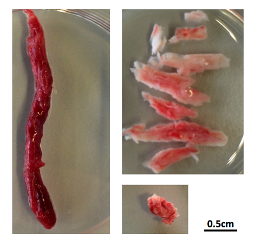
There are two main treatments for stroke caused by a clot in a blood vessel in the brain. One treatment, mechanical thrombectomy, involves pulling the clot out with a specialized catheter that is inserted into the artery in the groin and guided by imaging to the clot. This procedure is only performed at hospitals that specialize in these techniques. The other treatment, which is more widely accessible, involves giving a patient a clot-busting drug that helps the body dissolve the clot.
Quick decision making on which treatment is best for which patient is critical because the clot deprives brain cells of oxygen causing them to die. For physicians, knowing which patients will benefit the most from the clot-buster Alteplase (also known as tPA) just got easier.
University of Calgary scientists with the Hotchkiss Brain Institute at the Cumming School of Medicine (CSM) have discovered that clots have different compositions and depending on where they are located in the brain, administering tPA can be almost as effective as thrombectomy given sufficient time.
“We’ve known that, when administered quickly, tPA can be effective in stroke, but until now, we didn’t realize how effective it can be and we didn’t understand the specific reasons why it works better in some cases than others,” says Dr. Bijoy Menon, MD, associate professor in the departments of Clinical Neurosciences, Radiology and Community Health Sciences at the CSM. “Our findings show that some clots are permeable, which allows the tPA to penetrate the blockage and dissolve it. We saw that within two hours, greater than 50 per cent of permeable blockages had dissolved.”
The UCalgary study led out of the Foothills Medical Centre is the largest of its kind to date, involving nearly 600 patients at 12 medical centres in five countries (Canada, the Czech Republic, South Korea, Spain and Turkey). The findings are published in JAMA.
“Despite earlier research on the benefit of using tPA, we know there is still some reluctance in the medical community to use it. These findings should provide physicians with definitive evidence on the value of giving patients tPA as soon as they’ve confirmed the stroke is due to a clot,” says Dr. Andrew Demchuk, MD, professor in the departments of Clinical Neurosciences and Radiology. “It’s critical that anyone showing symptoms of a stroke be given a CT-angiogram as soon as possible to confirm the blockage. The scan will guide whether tPA is likely to dissolve the clot and may inform whether the patient also needs thrombectomy.”
A CT-angiogram (computer tomography scan) is a common noninvasive diagnostic tool that allows physicians to see images of the blood vessels in the brain. Researchers found that clots in the carotid artery of the brain do not respond to tPA, and for these patients, thrombectomy is required.
“Strokes happen at anytime, anywhere. Knowing who needs thrombectomy can help physicians make better decisions on how to prioritize patient transfers to specialized centres for this procedure,” says Menon. “Data gathered in Europe showed that up to one-third of hospital transfers aren’t necessary.”
“Stroke is an important health care problem and one of the leading causes of death and disability worldwide,” says Dr. Brian H. Rowe, scientific director, Canadian Institutes of Health Research (CIHR) Institute of Circulatory and Respiratory Health, which supported this study. “Through continued scientific research, important discoveries like this one will improve our ability to match patients with the most effective treatment for this particular injury. This will help speed up recovery times, reduce the associated impacts such as paralysis, and it will improve patient outcomes and ultimately save lives.”
Drs. Menon and Demchuk add that for the science community these findings will help researchers better design studies that target dissolving the clot with new clot busting drugs or combination treatments.
Led by the Hotchkiss Brain Institute, Brain and Mental Health is one of six strategic research themes guiding the university towards its Eyes High goals. The strategy provides a unifying direction for brain and mental health research at the university and positions researchers to unlock new discoveries and treatments for brain health in our community.
Story Source: University of Calgary. “Critical differences in clots that cause a stroke: Findings will help inform physicians which treatment will work best for patients.” ScienceDaily. ScienceDaily, 12 September 2018. <www.sciencedaily.com/releases/2018/09/180912081219.htm>.

Sep 4, 2018
The original article first published on ScienceDaily.com
About one third of patients who have suffered a stroke end up with low vision, losing up to half of their visual field. This partial blindness was long considered irreversible, but recent studies have shown that vision training after optic nerve and brain damage can help restore or improve vision. A new study published in the journal Clinical Neurophysiology reports on key mechanisms of vision restoration: attention.
Hemianopia is a decreased vision or blindness in half the visual field, usually as a consequence of stroke or trauma to the brain. It greatly reduces quality of life, affecting patients’ reading, driving and spatial navigation.
“Knowledge in this field is still rather fragmentary, but recent studies have shown that vision can be partially restored by vision training, which improves the deficient visual field sectors,” explains Prof. Bernhard Sabel, PhD, Director of the Institute of Medical Psychology at Magdeburg University, Germany, co-investigator of the study. “Neuroimaging evidence supports a possible role of attention in this vision restoration.”
The study confirmed this hypothesis by obtaining evidence from functional magnetic resonance imaging (fMRI) that visual training led to functional connectivity reorganization of the brain´s attentional network.
Seven chronic hemianopic patients with lesions of the visual cortex took part in vision rehabilitation training for five weeks. After the pre-tests all received training sessions lasting one and a half hours per day for six days per week for five weeks. Each training session, lasting about 60 minutes, was composed of six blocks with 120 training trials each, during which participants had to respond to specially designed visual stimuli on a computer monitor. The pre- and post-test included perimetry testing, contrast sensitivity testing and fMRI scanning one or two days before and after training, respectively. Each contrast sensitivity test consisted of 420 trials in six blocks. The visual rehabilitation training was performed with one eye open, which was randomly chosen, while the non-trained eye was covered with an opaque eye patch.
After training, the patients had significantly improved visual function at the training location, and fMRI showed that the training led to a strengthening of the cortical attentional network connections between the brain region of the right temporoparietal junction (rTPJ) and the insula and the anterior cingulate cortex (ACC).
“Our MRI results highlight the role of attention and the right TPJ activation as a component of vision restoration training in hemianopia,” notes lead investigator Yifeng Zhou, DSc, of the Hefei National Laboratory for Physical Sciences at Microscale and School of Life Science, University of Science and Technology of China, Hefei, P.R. China, and State Key Laboratory of Brain and Cognitive Science, Institute of Biophysics, Chinese Academy of Sciences, Beijing, P.R. China. “However, it is unclear whether the rehabilitation of attentional networks is the direct result of training or the result of the rebalancing of bottom-up sensory streams, which should be investigated in future studies.”
“This discovery that the brain´s attention network is a key mechanism in partially reversing blindness is an exciting advance in the field of restoring vision in the blind, and it opens up new avenues to design new therapies that are even more effective than current methods to help people with low vision or blindness,” concludes Prof. Sabel.
Story Source: Institute for Medical Psychology, Otto-v.-Guericke University Magdeburg. “Attention network plays key role in restoring vision after brain damage: New study highlights the role of attention as a component of vision restoration training in hemianopia.” ScienceDaily. ScienceDaily, 4 September 2018. <www.sciencedaily.com/releases/2018/09/180904114753.htm>.