
Nov 3, 2017
A new and exciting e-learning tool is being developed by SAFE to support Stroke Support Organisations around positive and effective stroke advocacy
The Stroke Alliance For Europe (SAFE) in partnership with the European Stroke Organisation (ESO) has begun to develop a new online e-learning tool for empowering stroke advocates from across Europe. The Stroke Support Organisation Faculty Tool (SSOFT) will help to build the capacity and capabilities of Stroke Support Organisations (SSOs) by developing their knowledge and understanding around how to build positive and effective advoacy campaigns for stroke prevention, diagnosis and treatment. (more…)
Nov 3, 2017
The 11th World Stroke Congress promises to attract acclaimed experts in stroke from around the world. The congress will showcase a cutting-edge educational and scientific experience, focusing on the latest developments in stroke prevention, acute management and restorative care after stroke.
This major Congress will be returning to North America for the first time in more than 12 years.
Canada has well-developed stroke care systems and has contributed to significant advances in stroke care through efforts in developing guidelines, clinical and health systems research. These efforts are ongoing and increasingly important in the face of an aging population and increased health care demands in Canada and around the world. (more…)
Nov 2, 2017
Researchers at Maastricht University Medical Center and Maastricht University have discovered why the brain is more sensitive to oxygen deprivation, or hypoxia, than other organs. Hypoxia caused by a stroke, for example, activates a specific mechanism that is protective in other organs but can be detrimental to the brain. ‘This discovery solves a long-standing mystery of the unique sensitivity of the brain to hypoxia,’ says head researcher and professor Harald Schmidt. The research results were published today in the leading scientific journal Proceedings of the National Academy of Sciences (PNAS). (more…)
Nov 1, 2017
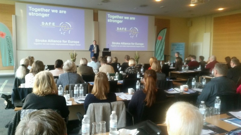
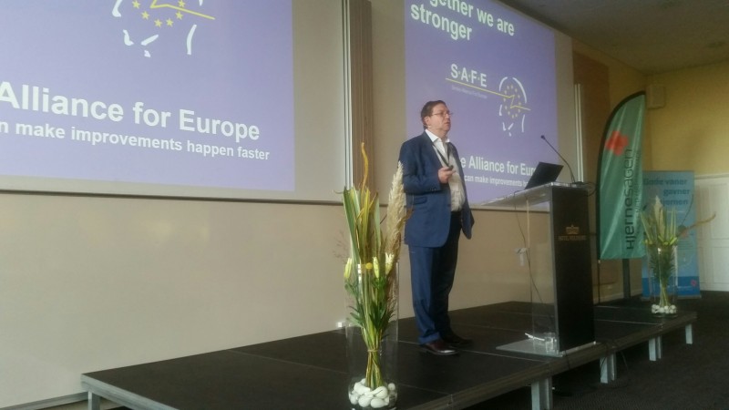
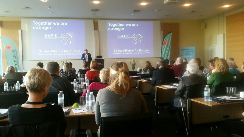
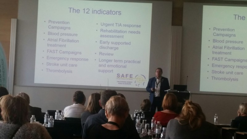
Research needs to be the platform for action in the decision making and policy sphere- said Jon Barrick, SAFE President at the World Stroke Day event in Denmark, on 30th October 2017.
Please see here the full presentation from this event.
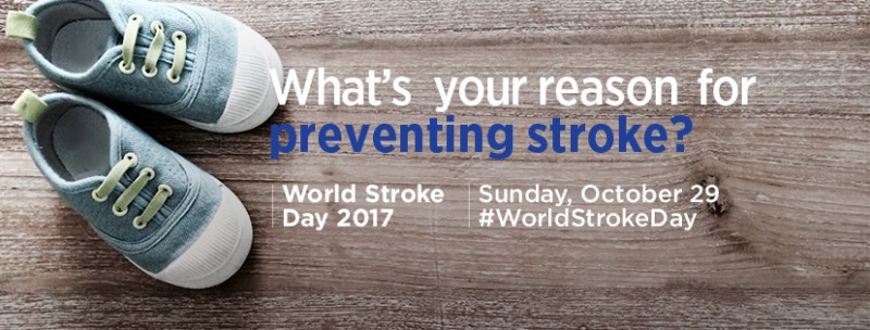
Oct 29, 2017
“WHAT IS YOUR REASON TO PREVENT STROKE?”
“One day we will be able to talk about a stroke and what it used to be and how we had a hand in stopping them.” – Brady Johnson, stroke survivor
Brussels, October 29, 2017: Good news is that stroke is preventable, with ten modifiable risk factors accounting for around 90% of the risk of stroke. Bad news is that we still largely fail to prevent it. On this World Stroke Day, we call upon Governments and health care system decision makers to implement population wide prevention strategies that address the biggest contributors to stroke- said Jon Barrick, SAFE President, adding that healthcare, researchers, stroke survivors and support organisations should all work together on developing and delivering effective national, regional and global stroke prevention strategies and campaigns. (more…)


